

Tel: (+86)15251899190
WeChat: KJ15251899190
E-mail: 2853036907@qq.com
Address: Complex building Lev11,
Jingang Sci-Tech Center, Nanjing.
Product Name: Ultrasound Transcranial Doppler Blood Flow Analyzer
Product model: KJ-2V6M
Detection principle: Ultrasonic Doppler principle
Domestic/Imported: Domestic
Medical/Household: Medical
Available for sale: nationwide
Input power: 60VA
Operating temperature: 5-40 ℃
Operating humidity: Relative humidity ≤ 80%
The following product introductions are all about the overall solution of the product
顱多普勒血流分析儀KJ-2V6M")
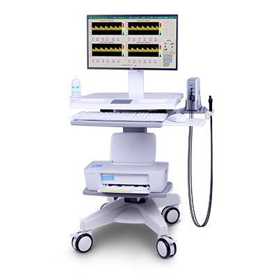
The KJ-2V6M model is a dual channel, four depth, and three probe ultrasound transcranial Doppler blood flow analyzer with three interface switching functions: single depth, double depth, and four depth. It can simultaneously observe the blood flow spectrum of multiple depths of a blood vessel, which has reference value for auxiliary screening of cerebral vascular stenosis. The overall solution of this product is equipped with a LCD computer all-in-one machine, a convenient trolley, and a color inkjet printer, Can conveniently achieve the detection function of ultrasound transcranial Doppler blood flow analyzer.
The KJ-2V6M model TCD instrument has M-mode and automatic blood vessel search functions, which can quickly search for blood vessels and greatly improve examination efficiency.
The KJ-2V6M model TCD instrument is equipped with two KJ-PW-2MHz and one KJ-CW-4MHz ultrasound probe, which can be used for intracranial vascular detection and extracranial vascular detection.
Functional characteristics
1. Ultrasonic probe technology
By using composite ceramic material chips, matching layer technology, and cutting processes, the signal-to-noise ratio and energy efficiency ratio of the sound beam have been improved, and the ultrasound penetration force and resolution of ultrasound images have been enhanced. Imitating the German probe structure, the signal-to-noise ratio and sensitivity have been improved.
2. Weak signal enhancement
Wavelet processing technology enhances weak signals, uses intelligent algorithms to analyze and process ultrasound echo signals, improves detection sensitivity, displays clear spectral images, and makes it easy to find vascular signals.
3. M module function
Equipped with M-mode function, it can simultaneously display multiple blood flow signals of different depths and quickly locate the depth of blood flow, helping doctors obtain blood flow spectrograms.
4. HITS detection technology
Equipped with embolus detection technology, when a embolus (HITS) appears, the signal displays a jumping waveform and can be replayed for recognition, making it easy for doctors to observe and detect.
5. Automatic vascular search function
The automatic blood vessel search technology captures weak signals, locates the depth of blood vessels, shortens the time to search for the location of blood vessels, and reduces the dependence on the operator's high level of technical skills.
6. Multi channel and multi depth detection
Simultaneously displaying multiple depths of a blood vessel, identifying typical symptoms such as vascular stenosis, and observing the trajectory of thrombus flow; Dual channel spectrum display comparison provides reliable evidence for bilateral vascular blood flow assessment.
7. Information management and transmission
Quickly record patient information, the software includes workstation functions, manage all patient information, and access patient testing data and reports at any time. DICOM 3.0 interface.
8. Other technologies
1. Dynamic and static envelope lines, real-time display of spectrum, calculation of blood flow parameters;
2. Multiple reports are available for easy modification;
3. Check medical records at any time, store vascular blood flow spectra for movie playback;
9. Clinical application
1. Used for blood flow measurement of transcranial, cervical, and peripheral blood vessels, excluding analytical and diagnostic functions;
2. Detect the blood flow of blood vessels in the brain;
3. Detection of blood flow within superficial blood vessels;
4. Quantitative evaluation of the blood supply status of cerebral and superficial vessels can provide a basis for screening the nature, degree, and scope of various cerebrovascular diseases.
Advice: Please carefully read the product manual or purchase and use it under the guidance of medical personnel. Please refer to the manual for any contraindications or precautions.
Tel: (+86)15251899190
WeChat: KJ15251899190
E-mail: 2853036907@qq.com
Address: Complex building Lev11,
Jingang Sci-Tech Center, Nanjing.
Copyright © 南京澳思泰生物科技有限公司 版權(quán)所有. All rights reserved. 蘇ICP備15032432號(hào)-1 網(wǎng)站地圖  蘇公網(wǎng)安備32011302321862